
NOMOSLOGIC
HYPOTHESIS PAPER · NOT YET TESTED · NO EXPERIMENTAL DATA

A Sulfenylation-Gated Lipid Peroxidation Relay at Mitochondria-Associated Membranes: A Cross-Field Hypothesis for Cancer-Selective Ferroptosis
Matthew Hardy · Founder and CEO, NomosLogic Inc
This is a hypothesis, not a finding. Nothing described in this piece has been observed in a laboratory. It is a mechanistic proposal, deliberately structured so that it can be proven wrong, with specific experiments, specific numeric thresholds, and named ways each part of the idea could fail. Anywhere this reads like a completed study, it is not one.
The Question This Hypothesis Tries to Answer
Ferroptosis, a form of regulated cell death driven by runaway oxidation of fats in cell membranes, kills cancer cells efficiently in a dish. The problem that has stalled its clinical use is selectivity: the same drugs that trigger ferroptosis in a tumor cell often trigger it just as well in the healthy cell next door. Existing explanations for the selectivity that does exist tend to be concentration arguments, how much GPX4 (the cell's main defense enzyme against this kind of damage) a given cell line happens to have, or how much of the antioxidant glutathione it has on hand.
This hypothesis proposes a different kind of variable: not how much protective enzyme a cell has, but where that enzyme is standing relative to where the damage is starting. It reframes ferroptosis susceptibility as a property of membrane geography rather than bulk biochemistry, and proposes a specific, nameable molecular reason why that geography would differ between a RAS-driven colorectal cancer cell and the normal colonocyte beside it.
The Core Hypothesis, In One Sentence
We hypothesize that a specific, reversible oxidation on a mitochondrial channel protein (VDAC2) acts as a geometric recruitment signal, not merely an on/off switch, that organizes a small patch of membrane next to itself: enriched in a fat species prone to oxidative damage, and locally depleted of the enzyme that would normally protect it. We further hypothesize that this patch persists specifically in RAS-driven cancer cells because the same oncogenic signaling that drives their growth also traps the repair enzyme that would otherwise dismantle the signal in the wrong compartment of the cell.
What Would Be New About This, If True
Six areas of biology already contain, individually, a piece of this picture. None of them, on their own, explain cancer selectivity. The novelty being proposed here is not any single fact, every node below is independently documented, but the specific sequence connecting them, which has not been assembled before.
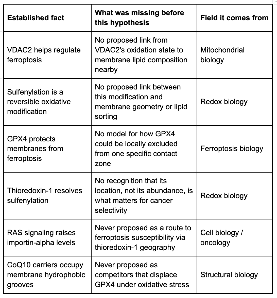
Why We Think This Requires Reading Six Fields Against Each Other
Redox biology alone can document that VDAC2 gets oxidized and that thioredoxin-1 resolves it, but has no framework for why that would matter spatially, or why it would be cell-type selective.
Membrane biophysics alone can model how charged surfaces sort lipids, but has no reason, on its own, to connect that to an upstream oxidative event or downstream cancer selectivity.
Structural biology alone can describe which residues on VDAC2 sit where, but cannot connect surface electrostatics to a lipid peroxidation outcome without the ferroptosis layer.
Cell biology and oncology alone know that RAS signaling raises importin-alpha and that thioredoxin-1 shows up in cancer cell nuclei, but have no mechanism tying that observation to a membrane-level ferroptosis vulnerability.
Ferroptosis biology alone knows that GPX4 loss drives lipid hydroperoxide accumulation, but has no molecular trigger explaining why any resulting vulnerability would concentrate at this particular membrane contact zone.
Mitochondrial biology alone can measure superoxide output and knows VDAC2 as a channel, but has no proposed link from the protein's oxidation state to downstream lipid sorting.
Remove any one field's contribution, in our view, and the proposed causal chain breaks. That is the basis for calling this a cross-field hypothesis rather than a restatement of any one field's existing findings.
The Idea in Plain Language
Cancer cells are, by several existing lines of evidence, unusually vulnerable to ferroptosis, their membranes can be made to oxidize themselves apart. The trouble is that healthy cells nearby can often be pushed into the same fate, which has limited ferroptosis as a therapeutic strategy. This hypothesis proposes a reason cancer cells might be secretly more vulnerable than their healthy neighbors, and, more specifically, proposes that the vulnerability is a side effect of the very mutation that makes the cell cancerous in the first place.
The Proposed Players
The mitochondria, the cell's power station
Every cell's mitochondria burn fuel for energy and, as exhaust, produce a reactive form of oxygen called superoxide. We hypothesize that cancer cells, which need to run their mitochondria harder to support rapid growth, produce meaningfully more of this exhaust than a healthy cell does under ordinary conditions.
VDAC2, a gatekeeper protein with two sensitive spots
VDAC2 sits on the mitochondrial outer wall and normally lets small molecules pass in and out. We propose it plays a second, unappreciated role: two of its amino acids (Cys77 and Cys232) act like a chemical mood ring, changing character when oxidative exhaust rises. We hypothesize that in a cancer cell's higher-exhaust environment, this mood ring gets stuck in its oxidized state far more often than in a healthy cell.
The membrane, a patchwork, not a plain sheet
The membrane around the mitochondria is not uniform; it is closer to a patchwork of different fat types. Two are relevant here: phosphatidylethanolamine (PE), a fat that is unusually easy to oxidize and, once oxidized, can pass the damage to its neighbors in a chain reaction; and phosphatidylserine (PS), a negatively charged fat that behaves differently at the same interface. We hypothesize that VDAC2's oxidized state changes the local electrical character of the membrane enough to pull PE toward it and push PS away, concentrating the most flammable fuel exactly where the sensor is stuck in its 'alarm' state.
GPX4, the fire-suppression enzyme
GPX4 is the only enzyme that can directly defuse oxidized PE inside an intact membrane, functioning like an embedded sprinkler system. We do not hypothesize that GPX4 disappears from the cell, only that it is locally crowded out of this one specific patch, by increased local traffic of a separate molecule (CoQ10) competing for the same membrane real estate.
15-LOX-1, the spark
This enzyme routinely oxidizes small amounts of PE as part of normal cell signaling; under ordinary conditions GPX4 catches each event before it spreads. We hypothesize that in the GPX4-sparse patch described above, this routine spark is left uncontained, and the oxidized PE begins oxidizing its neighbors.
CoQ10, the accidental crowd-out agent
CoQ10 shuttles electrons through the mitochondria's energy-production machinery. A cell running its mitochondria harder needs more of this traffic, and we hypothesize that traffic spills into the same membrane positions GPX4 would otherwise occupy, displacing it without inhibiting or degrading it.
Thioredoxin-1 (Trx1), the reset button
Trx1 can chemically reverse VDAC2's oxidized state. In a healthy cell, we hypothesize Trx1 sits near the mitochondria and performs this reset within minutes of each event, so the downstream lipid sorting and GPX4 displacement never have time to matter. If Trx1 is absent from that neighborhood, we hypothesize the oxidation, and everything that follows from it, persists indefinitely.
RAS and importin-alpha, the cancer cell's proposed fatal mistake
RAS is among the most frequently mutated genes in cancer, and constitutively active RAS is known to raise levels of importin-alpha, a protein that escorts other proteins into the nucleus. Trx1 carries a nuclear-targeting signal that importin-alpha recognizes. We hypothesize that in RAS-driven cancer cells, elevated importin-alpha continuously escorts Trx1 into the nucleus, where it has legitimate, separate work protecting nuclear DNA, leaving the cytoplasmic, mitochondria-proximal pool of Trx1 functionally depleted, not because less of it is made, but because too much of it is in the wrong room.
The Proposed Kill Sequence, Stage by Stage
Presented here as a hypothesized causal chain. Each stage is written in the language of prediction, and each is paired with the specific experiment we believe would test it (see the falsifiability section further below).
Stage 1: The cancer cell generates its own oxidative pressure
We hypothesize that ETC Complex I remodeling, a metabolic adaptation supporting rapid proliferation, elevates steady-state superoxide flux at the outer mitochondrial membrane in cancer cells, and that this is load-bearing for the cancer cell's survival, meaning it cannot simply be switched off.
Stage 2: VDAC2 Cys77 and Cys232 become sulfenylated
We hypothesize that the resulting hydrogen peroxide oxidizes these two surface-exposed cysteines to a sulfenic acid state at a much higher basal frequency in cancer cells than in normal colonocytes.
Stage 3: Sulfenylated VDAC2 electrostatically sorts PE to the contact point
We hypothesize that the partial negative charge introduced by this oxidation repels anionic PS headgroups and attracts zwitterionic PE headgroups directly at the VDAC2 contact point, without requiring a large conformational change in the protein.
Stage 4: GPX4 is displaced from the microdomain
We hypothesize that elevated CoQ10 carrier occupancy, itself a consequence of the cancer cell's harder-running mitochondria, competitively displaces GPX4 from the same hydrophobic membrane grooves, dropping local GPX4 concentration below the threshold needed for repair to outcompete propagation, without any change in total cellular GPX4 level.
Stage 5: 15-LOX-1 oxidizes PE without repair competition
We hypothesize that PE hydroperoxides accumulate rather than being neutralized, and that each one can oxidize an adjacent PE molecule, making the reaction self-sustaining once started, a chain reaction that would not require a continuous external oxidant.
Stage 6: The wave propagates via physical membrane continuity
We hypothesize that the reaction does not stay confined to its point of origin, but spreads laterally along the physical continuity between the mitochondria-associated membrane, the endoplasmic reticulum, and lipid droplets, the last of which represent a large reservoir of the same vulnerable fat species, potentially amplifying the signal considerably.
Stage 7: Ferroptotic membrane rupture
We hypothesize that accumulated lipid hydroperoxides across these connected membranes eventually compromise structural integrity, oxidized fat tails pack differently than native ones, and that iron-catalyzed Fenton chemistry accelerates the process further, ultimately reaching the threshold for plasma membrane rupture and regulated, non-apoptotic cell death.
Why We Propose the Healthy Neighbor Would Survive the Same Environment
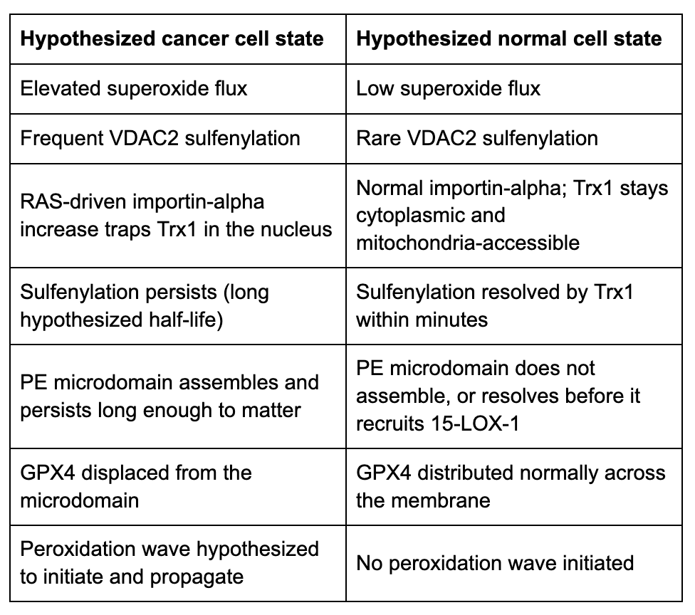
We propose the discriminating variable is not a binary switch but an integrated exposure, the product of how often sulfenylation happens (driven by superoxide flux) and how long each event lasts (driven by Trx1 accessibility). Cancer cells, in this framing, are hypothesized to have both a higher frequency and a longer duration; normal cells, both lower. The two cell types would not need to differ qualitatively in their underlying chemistry, only quantitatively, in kinetics, by a margin large enough to be therapeutically exploitable.
The Proposed Therapeutic Logic
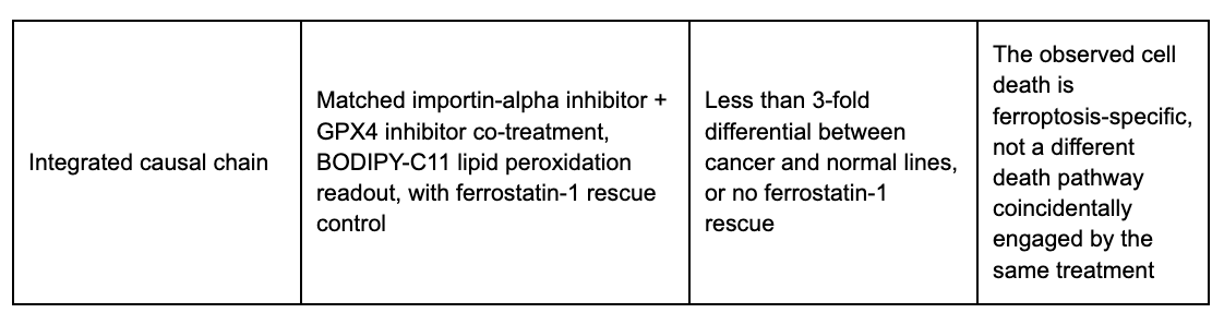
If the mechanism above is correct, we hypothesize that a two-drug combination could selectively push cancer cells across the ferroptotic threshold while leaving normal cells below it, not by inventing a new toxic mechanism, but by completing one the cancer cell has arguably already started on itself.
Proposed agent 1: an importin-alpha inhibitor
We hypothesize this would relocalize thioredoxin-1 back toward the cytoplasm in cancer cells, partially restoring VDAC2 resolution capacity. Used alone, we would expect this to modestly help the cancer cell, not harm it.
Proposed agent 2: a covalent GPX4 inhibitor
We hypothesize this would disable residual GPX4 activity everywhere in the cell. Used alone, existing evidence suggests this class of compound kills cancer and normal cells with similar efficiency, the therapeutic window is the open problem this hypothesis is trying to address, not one it assumes away.
The proposed combination
We hypothesize that in a cancer cell, where sulfenylation is already high and the PE microdomain already well established, even partially restored Trx1 would fail to fully reset the system before GPX4 inhibition removes the last line of defense, producing a comparatively fast ferroptotic response. In a normal cell, where the microdomain never assembled in the first place, we hypothesize that residual antioxidant capacity elsewhere in the membrane would be sufficient to prevent propagation even with GPX4 partially inhibited, leaving the cell under oxidative stress but below the threshold for rupture.
We want to be explicit that this combination has not been tested. Both proposed tool compounds have research-grade histories rather than clinical development records, and even if the underlying biology is confirmed, translating this into a clinical strategy would require substantial additional pharmacology work described in the research trajectory section below.
How We Would Know We Are Wrong
A hypothesis that cannot fail is not a hypothesis. Below is the experiment we believe would most cleanly test each mechanistic link, together with the specific threshold we believe the data would need to clear, and the single assumption whose failure would make the corresponding link uninterpretable rather than just weaker.
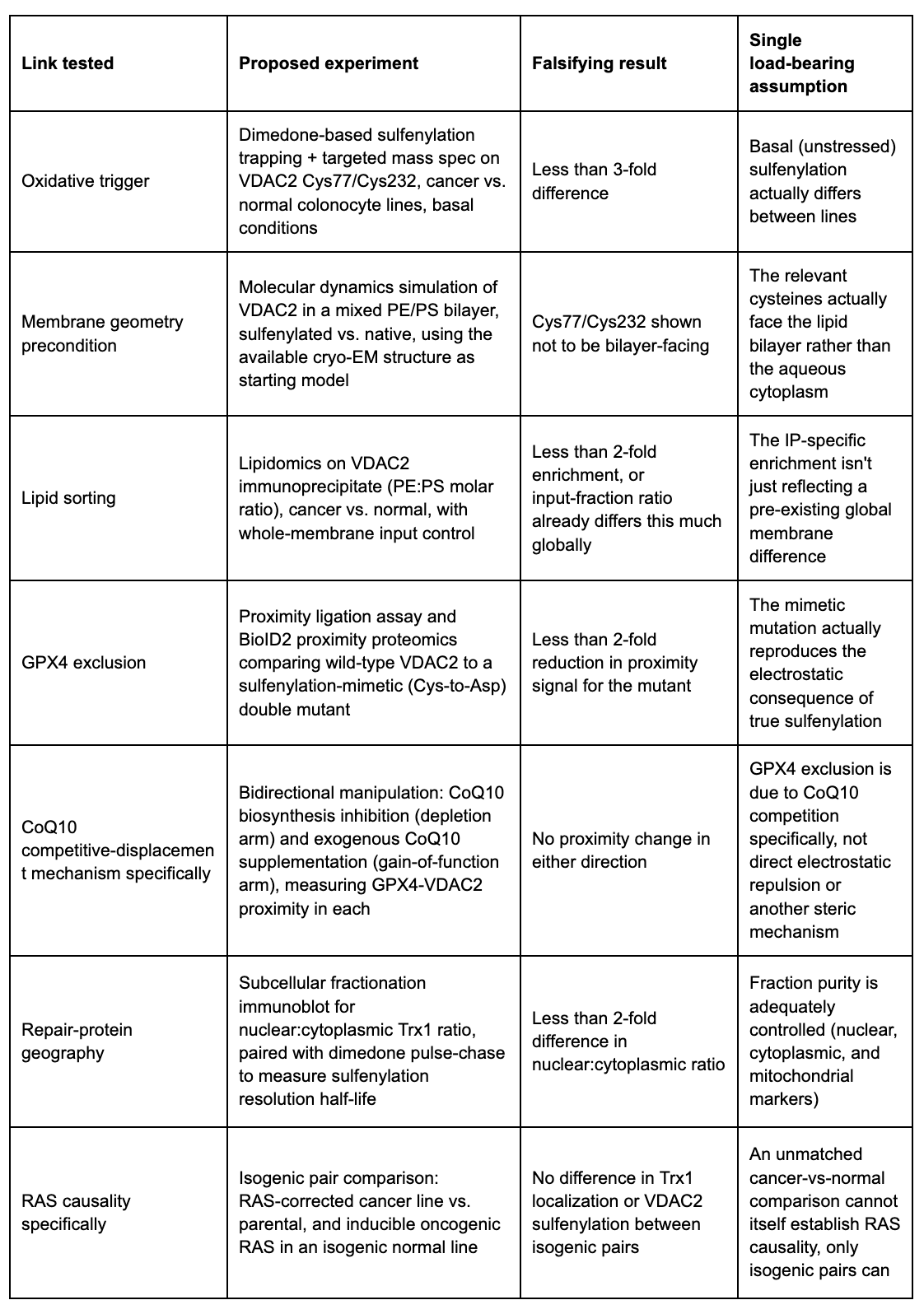
Proposed Gating Order
We believe these tests have a natural dependency order, and that running them out of order risks wasting effort on downstream experiments whose results would be uninterpretable if an upstream link fails:
1. Membrane-geometry simulation (a prerequisite check, answerable computationally before any wet-lab work)
2. Oxidative trigger experiment (cancer vs. normal, basal sulfenylation)
3. Isogenic RAS-pair check on Trx1 localization
4. Lipid-sorting experiment (PE:PS enrichment)
5. GPX4-exclusion experiment plus the CoQ10 bidirectional check
6. Combination therapeutic experiment, run last because it depends on every upstream link holding
Open Questions and Honest Limitations
The bilayer-facing topology of Cys77 and Cys232 has not been confirmed; the available structural reference was solved in detergent micelles, not a native membrane, so this is a genuine open question rather than a settled premise.
An unmatched comparison between one cancer line and one unrelated normal line cannot distinguish 'caused by the RAS mutation' from 'caused by some other difference between two unrelated cell lines.' Any claim of RAS causality requires isogenic pairs, not just a cancer-vs-normal comparison.
The CoQ10 competitive-displacement model is one of at least three plausible explanations for local GPX4 exclusion (the others being direct electrostatic repulsion or a separate steric/allosteric mechanism); it has not been distinguished from those alternatives.
This entire framework has, at most, been reasoned about in cell-line models on paper. Whether any part of it holds in primary tumor cells, organoids, or in vivo is unknown and would need to be established separately, in that order.
Whether other oncogenic drivers, or other cancer types, would show an equivalent Trx1 sequestration effect is untested; RAS mutation prevalence across cancer types makes this a plausible but unconfirmed extension.
Every numeric threshold in this piece (3-fold, 2-fold, and so on) is a pre-specified bar for a not-yet-run experiment. None of them are measurements.
Where This Would Lead, If the Early Gates Are Cleared
Immediate next questions
What is the physical size and lifetime of the proposed microdomain? Super-resolution imaging (STORM, PALM, or MINFLUX) could, in principle, directly visualize whether PE enrichment and GPX4 exclusion co-occur at the scale of a single VDAC2 molecule.
If the microdomain assembles and disassembles on a timescale of minutes, that would suggest a kinetic threshold model, potentially explaining why not every cancer cell with elevated VDAC2 sulfenylation undergoes spontaneous ferroptosis.
Medium-term extensions, if the core mechanism holds
Pan-cancer applicability
The RAS-importin-alpha-Trx1 axis is not inherently tissue-specific. If confirmed in colorectal cancer, it would be reasonable to ask whether the same vulnerability exists in pancreatic ductal adenocarcinoma (KRAS-mutant in the large majority of cases), non-small-cell lung cancer, or endometrial cancer, though this would need to be tested independently in each context, not assumed.
15-LOX-1 as a second biomarker
15-LOX-1 expression varies across colorectal cancer subtypes and is cytokine-regulated. If its activity turns out to be required for the proposed relay, then RAS mutation status and 15-LOX-1 expression together might define a more precisely stratified patient population, a hypothesis that could be evaluated in patient-derived organoids well before any clinical work.
Iron availability as a third axis
Ferroptosis is iron-dependent, and cancer cells often have larger labile iron pools. If that convergence turns out to matter here, it raises, but does not establish, the possibility of a three-way combination strategy, with a corresponding need for caution around iron-related toxicity in normal tissue.
Longer-term, more speculative questions
Is sulfenylation-gated lipid sorting a general membrane-organization principle, or specific to VDAC2? It is worth asking, if this holds, whether other oxidative modifications on other outer-mitochondrial-membrane proteins produce analogous effects.
Is spatially gated initiation specific to cancer, or a general feature of ferroptosis biology that simply operates at different thresholds in different physiological contexts (development, ischemia-reperfusion injury, immune cell function)?
Could the proposed microdomain, if directly imaged, serve as a pharmacodynamic biomarker, a way to confirm a treatment is engaging the intended mechanism in patient tissue, rather than inferring it indirectly?
What would be required before any clinical reasoning is appropriate
Confirmation in patient-derived organoid pairs (tumor and matched normal from the same patient), which is a materially more rigorous comparison than cell lines.
Development or identification of a clinically viable importin-alpha-pathway modulator, the tool compound referenced in this hypothesis has a research-only history, no established pharmacokinetics, and no clinical safety record.
A biomarker-stratified trial design, contingent on all of the above, is a downstream possibility to keep in view, not a near-term plan.
Why This Would Matter, If Confirmed
The prevailing question in this field has been: how much of a protective enzyme does a cell have? This hypothesis proposes a different question: where is that enzyme, relative to where damage is starting? If that reframing survives testing, it would suggest a class of therapeutic strategy built on spatial organization of repair machinery rather than its total abundance, and it would raise a broader question worth asking about other forms of regulated cell death: whether apoptosis, necroptosis, and pyroptosis carry similar spatial logic that bulk biochemistry has simply not been positioned to see.
Matthew Hardy is the CEO of NomosLogic, working at the intersection of computational drug discovery, translational biology, and AI-driven research infrastructure.
Tags: ferroptosis, VDAC2, sulfenylation, mitochondria-associated membrane, phosphatidylethanolamine, GPX4, thioredoxin-1, importin-alpha, RAS signaling, lipid peroxidation, membrane contact zones, colorectal cancer, redox biology, spatial biochemistry, falsifiability, cross-field hypothesis, cancer-selective cell death, drug discovery, nomoslogic,matthardy, drugdiscovery, research