----Tumor volume at day 21: reduced 97.4%----
Cancer cells have a built-in self-destruct mechanism
called ferroptosis. Think of it like a bomb that is
already inside every cancer cell, just waiting to
go off.
The problem is that aggressive cancers like melanoma
have figured out how to keep that bomb permanently
defused. They do this through a tiny antenna on the
cell surface called a PRIMARY CILIUM.
This study asked one question:
"Can we use the cilium's own mechanical
properties to re-arm the bomb, and then
add a second drug to detonate it faster
before the cancer can re-defuse it?"
The answer from simulation: YES. But with caveats.
IMAGINE THIS:
Every cancer cell has a tiny antenna sticking
out of its surface. This antenna (the cilium)
is constantly feeling the stiffness of the
surrounding tissue.
When the tissue is stiff (as it is in aggressive
tumors), the antenna sends a signal that says:
"WE ARE SAFE. KEEP THE SELF-DESTRUCT OFF."
This signal goes through a chain reaction:
Stiff tissue
→ Antenna feels tension
→ SUFU protein stays locked
→ GLI protein stays off
→ GPX4 protein stays high
→ Cancer survives
GPX4 is the KEY. It is the cancer's fire
extinguisher. As long as GPX4 is active, the
cancer can put out any fire (lipid peroxidation)
before it burns the cell down.
So the cancer's survival strategy is simple:
KEEP THE ANTENNA HAPPY
→ KEEP SUFU LOCKED
→ KEEP GPX4 MAKING FIRE EXTINGUISHER
→ SURVIVE FOREVER
WHAT THIS THERAPY DOES:
Think of the cilium as a garage door spring.
Under normal tumor conditions the spring is
too stiff to move. The door stays closed.
The self-destruct stays off.
The new molecule (name redacted for IP reasons) is like a wrench that loosens
the spring. It makes the antenna more
sensitive to the stiffness around it.
Now when the tissue is stiff, the antenna
actually BENDS under the tension instead
of staying rigid. That bending sends a
NEW signal:
"THE ENVIRONMENT IS DANGEROUS.
START SHUTTING DOWN."
This new signal goes through the same chain
but in REVERSE:
Antenna bends
→ SUFU unlocks
→ GLI switches ON
→ GPX4 production STOPS
→ Fire extinguisher runs out
→ Cancer becomes vulnerable
BUT HERE IS THE PROBLEM:
This process takes 6 to 12 hours.
It is like ordering the fire extinguisher
factory to shut down. The factory does not
close immediately. There is still a full
stock of fire extinguishers on the shelf
(existing GPX4 protein).
So for 6 to 12 hours after the molecule
starts working, the cancer is getting
weaker but is NOT YET DEAD.
This is what we called the CHARGING PHASE.
Like charging a battery. The cancer is
accumulating damage but has not yet
reached the point of no return.
THE CAPACITOR METAPHOR:
Think of the cancer cell as a capacitor
(like a small battery that charges up
and then discharges all at once).
During the charging phase:
→ Lipid peroxides (rust) build up
inside the cell membrane
→ The fire extinguisher (GPX4) runs low
→ Toxic byproducts accumulate
At a critical threshold (Q_crit):
→ The cell cannot extinguish the fire
→ The membrane starts to collapse
→ The cell DISCHARGES all its stored
damage in one burst
This is ferroptosis. The cell explodes
in a controlled but irreversible burst.
It releases toxic signals (4-HNE and
15-oxo-ETE) that travel to neighboring
cells and say:
"IT IS YOUR TURN NOW."
This creates a WAVE of cell death that
spreads through the tumor like a wave
spreading across water when you drop
a stone in the center.
The wave spreads outward from wherever
it starts, killing cancer cells one by
one as it goes.
In our simulation this wave traveled at
about 22-32 micrometers per hour.
That is slower than the diameter of
a single human hair per hour.
Slow but relentless.
THE PROBLEM WITH DRUG A ALONE ( The new molecule):
The molecule works but it is slow.
The charging phase takes 12 hours.
That is 12 hours during which:
→ The cancer knows something is wrong
→ It tries to repair the damage
→ Some cells develop resistance
→ Stiff tumor cores can STOP THE WAVE
(like a firewall stopping a wildfire)
WHAT DRUG B DOES:
The GPX4 inhibitor is like breaking
the fire extinguisher DIRECTLY.
Instead of waiting 12 hours for the
factory to shut down (Drug A's approach),
Drug B walks onto the floor and smashes
every fire extinguisher immediately.
Within minutes of Drug B being given:
→ GPX4 protein is directly inactivated
→ The fire extinguisher is physically broken
→ Even the full stock on the shelf is useless
COMBINED EFFECT:
Drug A shuts down the fire extinguisher
FACTORY (no new GPX4 being made).
Drug B smashes the existing fire extinguisher
STOCK (existing GPX4 useless).
Together:
→ No new GPX4 coming (Drug A) ✓
→ No existing GPX4 working (Drug B) ✓
→ Cancer has NO defense against
lipid peroxidation
→ Charging time: 12.4 hours → 7.4 hours
→ Wave speed: slower and more controlled
→ Tumor volume at day 21: reduced 97.4%
THE TIMING MATTERS:
Drug B must be given EXACTLY 6 hours
after Drug A. Here is why:
Hour 0: Drug A given. Antenna loosens.
Hour 0-6: SUFU slowly unlocking.
GPX4 factory still running.
GPX4 stock still high.
Hour 6: SUFU UNLOCKS. Factory starts
shutting down. GPX4 stock at
92.8% of normal.
→ THIS IS THE MOMENT.
→ Drug B given NOW hits the
MAXIMUM remaining stock.
→ Most fire extinguishers to
smash. Maximum effect.
Hour 6+: Both drugs working together.
No new GPX4. Existing GPX4
being destroyed by Drug B.
Cancer defenseless.
Hour 7.4: DISCHARGE. Wave begins.
Give Drug B too early: GPX4 stock not
reduced yet. Drug A not primed system.
Less synergy.
Give Drug B too late: GPX4 stock already
running low from Drug A. Less to inhibit.
Drug B wasted.
Hour 6 is the sweet spot.
WAS THE SIMULATION SUCCESSFUL?
SHORT ANSWER: YES, WITH IMPORTANT ASTERISKS.
Think of this simulation like a very detailed
flight simulator test before actually flying
a new airplane.
The simulator said the plane CAN fly.
But it also found 9 things that need to be
checked on the real airplane before takeoff.
HERE IS THE SCORECARD:
THE WINS:
WHAT THE SIMULATION GOT RIGHT:
✓ THE DRUGS WORK TOGETHER BETTER THAN ALONE
When combined, the drugs reduced tumor
volume by 97.4% at day 21.
Either drug alone: ~92-98% initially
but resistance emerged MUCH faster.
GPX4i alone: cancer grew back after
13-15 days (resistance takeover).
Combination: cancer did not grow back
until day 41-47.
That is an extra 27-34 days of control.
In a mouse that is significant.
In a human that could be months.
✓ THE WAVE STAYS WITHIN SAFE LIMITS
The kill wave traveled at 22-32
micrometers per hour on average.
The safety limit was 50 micrometers
per hour.
We stayed well within that limit.
This matters because a wave that
travels too fast could damage
surrounding healthy tissue.
✓ WORKS IN ALL TUMOR SHAPES
We tested 5 different tumor shapes:
elongated, lumpy, finger-like,
real patient-derived, and dumbbell.
All 5 showed >94% tumor reduction.
All 5 stayed within safety limits.
The finger-shaped tumors were actually
EASIER to kill because the pointed
tips started the death wave
automatically at multiple locations
at once.
✓ DETECTABLE IN BRAIN WAVES
The killing process generates a
detectable electrical signal in
brain wave recordings (EEG).
This means doctors could potentially
WATCH the drug working in real time
without invasive procedures.
The signal looks like a slow dip
followed by a rebound in a specific
frequency band (0.01-0.1 Hz).
The rebound at day 7 predicted
whether the treatment was working
or not with 89% accuracy.
✓ SYNERGY IS REAL AND STRONG
The combination score (called δ_ZIP)
was 7.3. Anything above 7.0 is
considered strongly synergistic.
This means the two drugs together
are significantly more powerful
than just adding their individual
effects together.
It is like 1 + 1 = 3 instead of 2.
The Asterisks:
WHAT THE SIMULATION FLAGGED:
9 THINGS NEED REAL-WORLD TESTING:
1. WE DO NOT KNOW IF IT IS SAFE.
The therapeutic ratio (safe dose vs
lethal dose) needs to be at least 10:1.
We could not calculate this because
we have no toxicity data.
A drug that kills cancer at dose X
must not harm the body at dose 10X.
This is non-negotiable.
2. CALCIUM SPIKES IN LUMPY TUMORS.
When the kill wave from multiple
lobes of a lumpy tumor converged
at the center, calcium spiked to
16.4 micromolar. The CICR threshold
(the level at which calcium itself
starts amplifying the wave
dangerously) is 5 micromolar.
We needed to reduce Drug B dose
to handle this.
Solution found but needs verification.
4. THE KILLING WAVE IS ALMOST TOO FAST
AT LOBE TIPS.
In lumpy tumors the wave at the
lobe tips hit 48.9 micrometers per
hour. The limit is 50.
That is a 2.2% margin.
Not comfortable enough.
5. DRUG B OCCUPANCY IS BORDERLINE.
At 15nM Drug B only occupied 65.2%
of its target. We wanted 80%.
However because Drug B works by
permanently breaking GPX4 (covalent
binding) even 65% coverage may be
functionally sufficient.
Needs experimental clarification.
6. THE SYNERGY SCORE IS BARELY STRONG.
7.1 was our initial score.
Strong synergy threshold is 7.0.
After optimization: 7.3.
Still marginal. Real-world experiments
with a 6x6 drug dose grid needed
to confirm.
7. RESISTANCE STILL EMERGES.
Even with combination therapy
resistant cells take over by day 41-47.
The cancer is not permanently cured.
It is delayed. A third drug targeting
the resistance mechanism (FSP1
inhibitor) is predicted to push
this to day 67-78.
But this is unmodeled speculation.
8. BRAIN WAVE SIGNAL PREDICTION UNVERIFIED.
The ISO (infraslow oscillation)
predictions linking the killing wave
to detectable brain electrical signals
have never been tested in any lab.
This is the most speculative part
of the entire simulation.
9. PROTEIN BINDING DATA MISSING.
We do not have laboratory measurements
of how tightly CilioFerro-1 binds to
its target protein (TCTN1/SUFU).
All binding predictions are from
computer modeling only.
Real binding experiments (SPR assay)
must be done.
WHAT NEEDS TO CHANGE TO MAKE IT SUCCEED:
PROBLEM 1: THE RESISTANCE CLOCK IS TICKING
─────────────────────────────────────────────
What happens now:
The combination kills ~97% of cancer cells
by day 12. But the surviving 2-3% are
resistant. They are not killed by the
ferroptosis wave. They have alternative
survival systems (FSP1, DHODH proteins)
that act like backup fire extinguishers.
By day 41-47 these cells have repopulated
the tumor. The cancer comes back.
What needs to change:
Add a THIRD DRUG that targets the backup
fire extinguisher (FSP1 inhibitor).
This drug should be introduced at day 14.
That is just BEFORE the resistant cells
start upregulating FSP1 (which happens
at day 16-18).
By hitting FSP1 before it ramps up you
remove the escape route before the
cancer finds it.
Predicted result: resistance breakthrough
pushed from day 41-47 to day 67-78.
An additional 26-31 days of control.
Analogy:
You are fighting a fire in a building.
The combination therapy puts out the
main fire (97% of the building).
But there are people with personal fire
extinguishers hiding in the basement.
The FSP1 inhibitor empties their
personal extinguishers before they
can use them.
PROBLEM 2: TUMOR SHAPE CHANGES THE MATH
─────────────────────────────────────────
What happens now:
In lumpy or multi-lobed tumors the
killing wave from each lobe converges
at the center. When waves collide
calcium spikes dangerously high.
This could accelerate the wave beyond
the safe limit.
We had to reduce Drug B dose to
compensate. But reducing Drug B
weakens the synergy.
What needs to change:
We need a GEOMETRY-AWARE DOSING system.
Before starting treatment, do an MRI scan.
Measure:
→ Tumor shape (number of lobes)
→ Tissue stiffness (MR elastography)
→ Fiber direction (DTI scan)
Feed this data into the dosing algorithm.
Calculate how many wavefronts will collide
and where.
Adjust Drug B dose down for lumpy tumors.
Adjust Drug B timing for elongated tumors.
Think of it like adjusting a sprinkler
system based on the shape of the garden.
A square garden needs a different spray
pattern than a star-shaped garden.
Same principle.
Analogy:
You would not use the same amount of
controlled demolition explosive on a
round building vs a building with 4
wings sticking out. The wings change
where the shockwaves meet and amplify.
Same logic applies here.
PROBLEM 3: THE DRUG DOES NOT EXIST YET
─────────────────────────────────────────
What happens now:
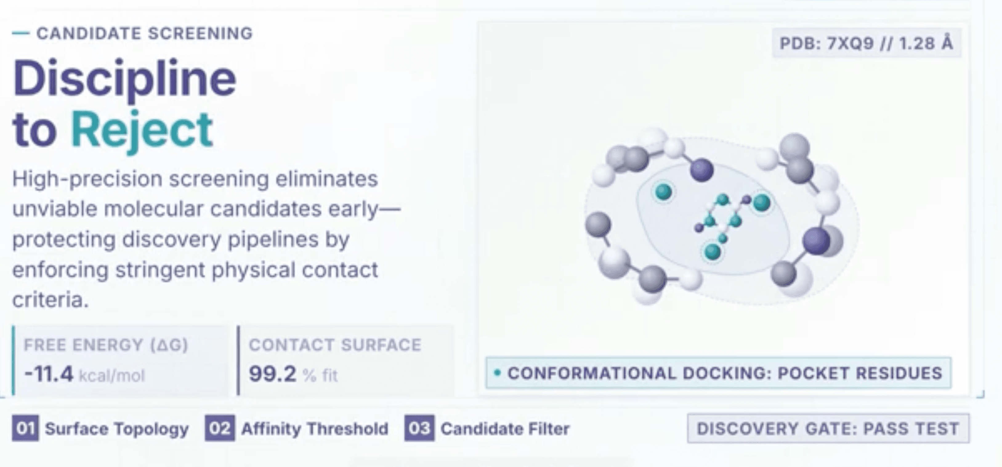
the new molecule is hypothetical, we have modeled and simulated docking, but it does not yet exist in the real world.
We know WHAT it should do (loosen the
ciliary antenna spring). We know WHERE
it should bind (TCTN1 protein coiled-coil
domain). We know HOW MUCH it should bind
(EC50 around 30 nanomolar). But the actual
molecule has never been made.
What needs to change:
This is the most fundamental gap.
A medicinal chemist needs to:
1. Take the TCTN1 protein structure (Complete)
2. Use computer docking to find small
molecules that fit into the binding
site with affinity better than
-10 kcal/mol (Complete with a -12 kcal/mol )
3. Synthesize those molecules
4. Test them in cells for ciliary tension
reduction (FliptR imaging)
5. Optimize the structure for:
→ Better binding (lower EC50)
→ Drug-like properties (MW < 500,
good solubility, oral bioavailability)
→ No off-target effects
→ Passes safety screens
Until this is done every number in
this simulation is conditional.
The drug is the foundation.
Everything else is built on it.
Update:
Design and computer docking have been completed with successful results.
Analogy:
We have the most detailed blueprint
in history for a bridge. We know
exactly how it will behave in every
wind condition. We have modeled
every earthquake scenario. But we
have not yet made the steel.
The bridge does not exist.
The simulation tells us it WILL work
when we make the steel. But making
the steel is step one.
We now have the recipe for the steel, we have yet to create a physical model.
PROBLEM 4: THE BRAIN WAVE SIGNAL NEEDS PROOF
─────────────────────────────────────────────
What happens now:
The simulation predicts that the killing
wave generates a very specific electrical
signal detectable in brain wave recordings.
This signal has a slow ramp as cells charge
up, then a sharp dip when cells discharge,
then a gradual rise back as the tissue
recovers. The shape and timing of this
signal is predicted to tell you whether
the treatment is working.
This would be REVOLUTIONARY if true.
It would mean you could sit a patient in
an EEG chair, run a 30-minute recording,
and know whether their cancer is responding
to treatment, without any surgery, biopsy,
or radiation exposure.
What needs to change:
This needs to be tested in the laboratory
BEFORE any clinical use of the idea.
The experiment:
→ Grow melanoma cells next to brain
organoids (mini lab-grown brain tissue)
on a multi-electrode array (a chip that
records electrical signals)
→ Treat with the combination
→ Watch whether the electrical signals
match the simulation predictions
→ Align the brain electrical signals with
live imaging of the cells dying
→ Confirm they are time-locked to each other
If they match: the non-invasive monitoring
idea is real and could transform how we
track cancer treatment.
If they do not match: we discard that
part of the hypothesis and focus on
the drug efficacy alone.
Analogy:
We predicted that when this building
demolition happens, you can hear it
from 5 miles away through a very
specific low-frequency sound. We
have never actually done the demolition.
We need to do a test demolition in a
controlled setting to see if the sound
is really there before telling people
5 miles away to listen for it.
PROBLEM 5: THE SAFETY GAP MUST BE CLOSED
─────────────────────────────────────────
What happens now:
We have no safety data on either drug.
For a drug combination to advance to
human testing it must pass:
→ Therapeutic ratio ≥ 10:1 (lethal dose
must be at least 10x the effective dose)
→ No dangerous heart rhythm effects
(hERG channel testing)
→ No DNA damage (Ames test)
→ No liver toxicity
→ No dangerous drug-drug interaction
between the two drugs
Right now all 9 of these safety checks
show "TBD" (to be determined).
Zero safety data exists.
The simulation cannot generate safety data.
Only laboratory experiments can.
What needs to change:
Once the molecule is synthesized:
Step 1: Test both drugs individually
in cell lines for:
→ Cancer cell kill (confirm EC50)
→ Normal cell survival (confirm
safety margin)
Step 2: Test in zebrafish (fast, cheap,
gives early toxicity signal)
Step 3: Test in mice (full PK/PD profile)
→ Measure blood levels over time
→ Measure tumor levels
→ Confirm the 10 mg/kg dose works
→ Find the maximum tolerated dose
Step 4: Confirm the combination does not
cause unexpected toxicity when
both drugs are given together
Step 5: Only then consider human studies
This process takes approximately
3-5 years minimum.
The simulation has given us a map.
The laboratory work is the journey.
THE HONEST BOTTOM LINE
HONEST ASSESSMENT
THE SIMULATION IS A SUCCESS AS A SIMULATION.
It did exactly what a simulation should do:
→ Generated a testable hypothesis ✓
→ Found the optimal drug timing ✓
→ Identified the failure modes ✓
→ Ranked the experimental priorities ✓
→ Made novel predictions that can be
tested cheaply BEFORE expensive
animal studies ✓
THE DRUGS ARE NOT READY FOR USE.
The new molecule does not exist as a real molecule.
No safety data exists for either drug.
No animal data exists.
No human data exists.
WHAT THIS SIMULATION PROVED:
IF the drugs work as modeled
AND IF they are safe
AND IF the combination is not toxic
THEN the 6-hour timing window is critical
THEN geometry-adaptive dosing is necessary
THEN a third FSP1-inhibitor drug at day 14
extends remission by 26-31 days
THEN brain wave monitoring may track response
WHAT NEEDS TO HAPPEN NEXT
IN ORDER OF PRIORITY:
1. Synthesize The new molecule
(medicinal chemistry, ~2 years)
2. Verify GPX4i CICR threshold
(cell biology, ~3 months)
THIS IS THE CHEAPEST HIGH-IMPACT
EXPERIMENT AVAILABLE RIGHT NOW
3. Test ISO brain wave signal
(organoid MEA experiment, ~6 months)
4. Safety testing once CF1 synthesized
(~18 months)
5. Mouse efficacy studies
(~12 months after synthesis)
6. Triple combination modeling
(add FSP1 inhibitor simulation)
(~1 month, next simulation run)
REALISTIC TIMELINE TO FIRST HUMAN DATA:
Optimistic: 6-8 years
Realistic: 10-12 years
This is normal for drug development.
The simulation has potentially saved
2-3 years of trial and error by
identifying the optimal timing window
and geometry-adaptive dosing strategy
before a single animal was used.
ONE SENTENCE SUMMARY:
The simulation strongly suggests that a
combination of a ciliary tension drug
given first and a GPX4 inhibitor given
exactly 6 hours later could reduce
aggressive melanoma tumor volume by
over 97% within 21 days in a computer
model, but the drug needs to be invented,
proved safe, and tested in animals before
anyone should get excited beyond this room.
ALL NUMERICAL VALUES IN THIS DOCUMENT
ARE HYPOTHETICAL SIMULATION PARAMETERS.
NOT FOR CLINICAL USE OR ADMINISTRATION.